What is a Muscle Cell?
Our bodies are predominately made up of millions of muscle cells that make up our more than 640 individual muscles. Muscle cells are our body’s engine that allows us to function and survive. Without muscle cells we would not be able to move or have any ability to exist. Our muscles are our “metabolic engine” (no different from the car’s engine) that controls every aspect of our active life.
Muscle Cell Structure:
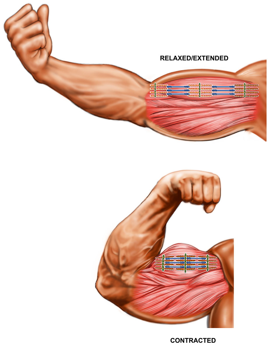
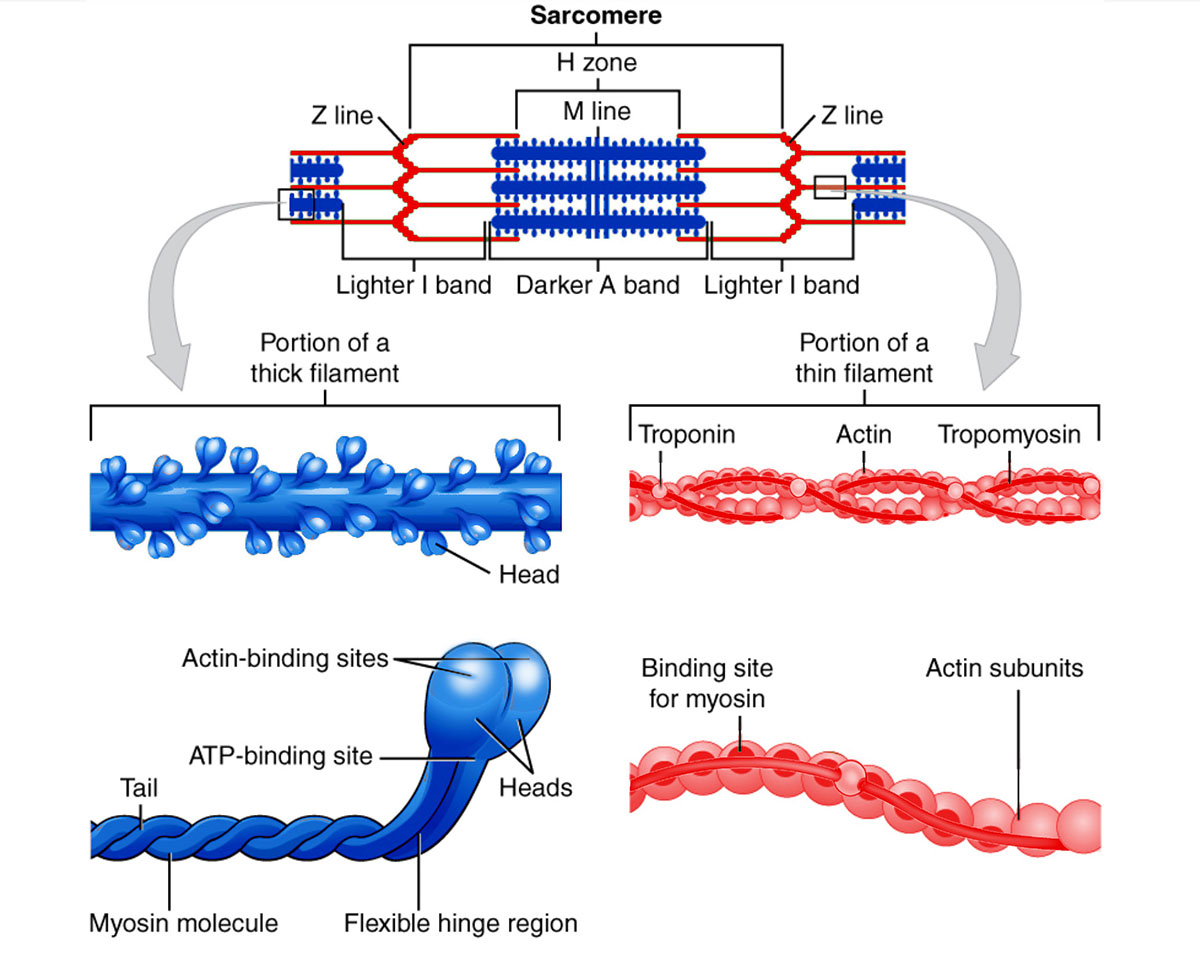
- ACTIN and MYOSIN: A muscle cell is long, round, and tubular in shape. Inside each muscle cell is a series of specially arranged motor proteins called actin and myosin, (made up from amino acids) which consist of thin and thick filaments that slide past each other, shortening the cell’s length and causing the cell to contract when required to perform a muscular task/movement.
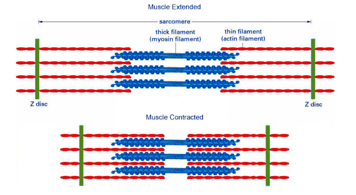
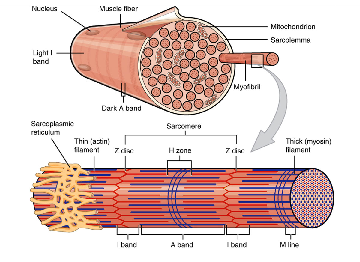
- SARCOMERES: Zooming into a single muscle cell, the cell is long and thin. That cell is segmented, organized by structures called sarcomeres. Each sarcomere contains myosin and actin. To visualize, if you have a 6-metre tube and you cut the tube into six pieces, the six pieces would be the sarcomeres. They are a sequential structure, one after the other, that form the length of the tube/muscle cell. They are defined at either end, like a square is defined by its edges, by two disks. These are structural proteins that scaffold the ends of each sarcomere. They are like pillars that give the sarcomere structure. Between each sarcomere is the “meat” of the muscle components-the actin proteins are anchored to the sarcomeres and jut inwards perpendicular to the sarcomere. Also attached, but loosely by a “spring-like” protein, is myosin. Myosin proteins are attached to a spring-like protein that has the ability to extend or squeeze together, allowing myosin to move, although it is still indirectly attached to the structural Z disk of the sarcomere.
- MYOFIBRILS: Each muscle cell contains thousands of sarcomeres, which make up a larger muscle fibre called myofibrils. Muscle cells contain densely compacted bundles of myofibrils which contain many sarcomeres that are bundled together. Many muscle cells connect together to form long fibers in muscle tissue. They are aligned parallel to one another like stacking tubes that attach to the bone and one another my tendons and collagen.
Mitochondria:
Each muscle cell contains hundreds of mitochondria. They are a muscle cells power plant that converts glucose and oxygen into the energy molecule ATP. This energy produced by the mitochondria is important during intense exercise when energy requirements by a muscle cell rapidly increases. It has been found in studies that energy produced by the mitochondria gets distributed among interconnected mitochondria within the same muscle cell through a series of fusion/connections. “They found that the mitochondrial “wires” were electrically conducive and that most of the mitochondria were in direct electrical communication through the interconnecting network exchanging different molecules between each other”. The process of using oxygen to convert glucose to ATP is called Oxidative phosphorylation. This process also uses oxygen to convert fatty acids and glycerol into ATP.
Nuclei (Nucleus):
The nuclei is the brain of the muscle cell because it is the main control center. It contains most of the DNA genetic material in our cells. Meaning they contain the blueprints for production of all proteins or cell needs. So, when a muscle cell is damaged by hi-intensity exercise, specific proteins that are damaged are removed through the cleanup machinery (proteasome, autophagy) and then replaced by new identical proteins. They contain the blueprints for production of all cell proteins or cell needs. So, when muscle is damaged by exercise, specific proteins that are damaged are removed through the cleanup machinery and then replaced by new identical proteins. The production of these proteins begins in the nucleus.
Signalling proteins known as transcription factors will move from the cytosol to inside the nucleus where they will bind to genes that hold the information for the proteins of interest. This will lead other proteins that are in charge of creating replicas of the information to bind and begin generating these replicas (known as RNA). The RNA is then moved out of the nucleus, where proteins bind to it and begin reading the RNA replicas of the gene. When they are read they stitch together the amino acids to create the specific new proteins. The bottom line is that without the genetic material/genes in the nuclei, there would be no muscle repair.
Sarcoplasmic Reticulum:
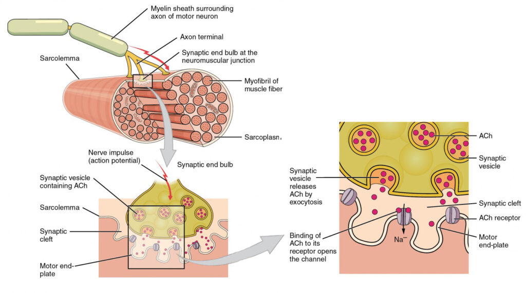
The sarcoplasmic reticulum is a large structure on the inside of the cell membrane which contains large stores of calcium. When the muscle is activated to contract by the central nervous system‘s (CNS) neuron , that attaches to the cell membrane, the sarcoplasmic reticulum releases its calcium into the cytosol of the cell. Once there, the calcium will fall into the myofibrils (the proteins made of actin and myosin among other contractile proteins) and bind to the troponin protein. The troponin protein sits on top of the tropomyosin protein-which sits on top of the actin filaments. Tropomyosin wraps itself around actin when the muscle is at rest so that myosin cannot grab on to its binding site. (Keeps myosin and actin away from each other so that they do not contract). When the calcium binds to troponin it shifts troponin and tropomyosin away from the actin, allowing myosin to bind to actin and begin muscle contractions. When a muscle is tired or overtrained, it doesn’t feel like it is contracting with any great force. This is because the nerve cell connected to those fibres are overworked and therefore transmission through the central nervous system is hindered.
The muscle has ways of communicating back to the spinal cord through sensory systems, as well as by chemical means in the sarcoplasmic reticulum, inhibiting the function of the activating neuron. Basically you have an afferent neuron (a single nerve cell) that protrudes from the spinal cord that innervates (communicates) with the muscle cell. When told to contract, the afferent neurons will activate the sarcoplasmic reticulum and contraction will occur. However, within the entire muscle there are millions of muscle cells that make up the biceps muscle (for example), there is feedback that is also communicated back to the spinal cord through other neurons knowing as efferent sensory neurons. If these neurons get triggered, they will override the efferent neuron at the spinal cord, inhibiting muscle contraction on a cellular scale. Alternatively, the muscle can also send out chemical signals through it’s efferent neurons and inhibit it as well.
What is Cytosol?
Cytosol is the area within a muscle cell that is not occupied by other key “factories/organelles, like the mitochondria or the sarcoplasmic reticulum”
Muscle cells store glycogen and water in the cytosol.
Muscle Cell Function
- CONTRACTION: If muscles are at rest, the muscle cells are at rest – meaning, molecularly, that the actin and myosin proteins that are parallel to one another inside the sarcomere are not interacting. They are stationary. However, the moment a person puts weight/load on the muscle, the muscle cell must react, so the myosin reaches over and attaches to the actin. The muscle cell has now established resistance, because what would have freely slid back and forth (because no interaction between myosin and actin) is now attached and holding firm. If more weight/load is thrown on the muscle, more actin and myosin proteins must interact to hold the weight/load in place. To move the weight/load against gravity (from an isotonic contraction to a concentric contraction), the myosin must crawl up the actin, shortening the muscle fiber (as the sarcomeres pull together, the Z disks get closer to one another, shortening this “dynamic pipe”).
Role of Satellite/Stem Cells in Exercised/Microscopic Damaged Muscle Cells:
Skeletal Muscle:
Skeletal muscles are attached to our ligaments and bones and have to be stimulated by nerve impulses to contract. These muscles literally cover our skeletons, form our muscular system, keep our posture, stabilize our joints, and give us our body shape. Skeletal muscles are our “metabolic engine” which generates heat, expends calories and helps us to maintain a functional body temperature.
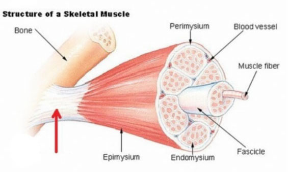
Our skeletal muscles come in all shapes and sizes and are attached to our bones directly or by other connective tissues called tendons. Skeletal musculature forms approximately 35% to 50% of our total body weight, making them the strongest part of our anatomy. Our muscular system forms our active motor system.
How Skeletal Muscles Work:
The job of skeletal muscles is to perform the movements of our limbs and body parts we asked them to perform as we go about our day-to-day activities. In other words skeletal muscles respond to voluntary control. You “tell“ them what to do by sending them messages from your brain to your muscle cells. Nerves carry electrical signals from the brain through the spinal cord’s central nervous system, (CNS) to each muscle in order to direct and control their movement. This impulse commands them to contract (shorten by contracting) by the nerve endings releasing chemicals into the muscle cell, which the muscle cell translates to contract and pull on our tendons. This moves the bones, thereby allowing us to move around.
During exercise, muscle fibres contract to the fullest extent, not just partially – a fact that is the basis of what’s called the “all or none” law of muscle physiology.
This does not mean that all the muscle cells/fibers contract when movement occurs, but the fibers that do contract do so to their fullest capacity. The demand placed on the muscle determines how many muscle fibres are called into action to contract simultaneously and complete the task. The more difficult the task or exercise, the more muscle fibers are activated. HIT3 training system’s main emphasis is to recruit and induce growth stimulation on the maximal amount of muscle fibres with each repetition performed-of every set of exercise.
Our bodies expend the least amount of effort possible to complete a task. When we exercise, in addition to the direct muscle fibres needed for the task, other stabilizing muscle fibres may become involved indirectly, but are not stimulated in a major way.
The muscles in our body are arranged in groups that usually work in pairs to perform opposite movements. An example is the bicep, or the upper arm, which we use to lift something up (by bending the elbow). This muscle has behind it the triceps, which we engage when we push something down (to straighten the elbow).
Whenever we exercise, whether it be weight training, swimming, running or bicycling, we perform repeated, coordinated movements that originate in our brains. As we gain experience doing exercises, we get better and better at them because our brains learn to send the messages more efficiently, as skills improve. Just like learning to ride a bicycle, exercise movements at first may feel a bit awkward or uncoordinated, but after a few sessions, movement will feel much smoother and productive.
Muscle Cells Energy Production:
Glucose enters a muscle cell membrane with the help of insulin, through tunnels called GLUT transporters. Glucose that is not immediately needed for energy is tagged and stored in the Cytosol chained together and called glycogen, (which are glucose molecules bonded together). A muscle has 4 distinct processes/pathways that produce energy by converting glucose or fatty acids to ATP.
Glycolysis:
Takes place in the cytosol of the muscle cell. Glycolysis only uses glucose molecules. When glucose enters the muscle cell, glycolysis will alter the structure of the glucose molecules, and doing so it will generate ATP (cellular energy). In the case when it is using stored glycogen, it will untag glycogen into glucose molecules (with enzymes) to produce ATP for immediate use.
Now glycolysis is broken into two types – aerobic and anaerobic
- Anaerobic Glycolysis:
In anaerobic glycolysis, glucose is used without oxygen present. In this case, glycolysis generates ATP as the glucose goes through that system; however, at the end, the final molecule produced is called lactic acid (lactate with an hydrogen atom). For HIT3 hi-intensity exercise, anaerobic glycolysis produces an extreme amount of lactic acid in the muscle cell. It is the hydrogen atom that causes the burn. After completing the exercise, during rest/recuperation, the lactate and hydrogen is shunted away from the muscle cell out to the blood stream to be metabolized in the liver.
- Aerobic Glycolysis:
Is glycolysis with oxygen present. In this case glycolysis uses glucose and oxygen to generate a molecule called pyruvate. The pyruvate molecule then goes to the mitochondria to generate ATP for aerobic metabolism-low level or moderate intensity work. Unlike lactate, pyruvate is not shuttled out of the cell. So both forms of glycolysis generate ATP. They do so quickly, but aerobic glycolysis will generate more energy so long as oxygen is present.
- Oxidative Phosphorylation:
Produces energy in 2 ways:
It takes a molecule of oxygen and pyruvate from the mitochondria and produces more energy for low intensity work and exercise. It also takes fatty acids from the cytosol to the Mitochondria and with oxygen produce ATP
- Phosphocreatine System:
Is based around the creatine molecule. The muscle cell largely operates on ATP for energy, but when ATP is used it turns to ADP. So, a fully charged cell contains ATP and a cell that has used its energy (ATP), will experience a rise in spent ATP, known as ADP. Creatine plays into this by holding on to phosphate molecules – creating the phosphocreatine molecule (Creatine + phosphate). When ADP levels begin to rise in the cell, the phosphocreatine will donate its phosphate to the ADP, recycling it into ATP. So you can think of phosphocreatine as a buffer from the cell getting into low energy state (ADP).
For greater clarity, the phosphocreatine system provides ATP to a muscle cell when the cell is deficient in glycolysis anaerobic ATP. This system is short-lived and can only supply energy for 10 to 15 seconds per exercise.
Bodybuilders take creatine for the purpose of supplying muscle cells more creatine molecules with phosphate that allows for the potential for the muscle to recycle the ADP into ATP for immediate energy. However, if the creatine is not taken continuously for days on end, it will not build up in the muscle cell to help ATP development. A muscle loaded with creatine will force the muscle cell to retain more water in order to maintain its dilution concentration. The bottom line is that no real muscle is built by taking creatine.
How do muscles get energy when glucose/glycogen is depleted during the performance of hi-intensity exercise?
Muscles depleted of glucose/glycogen have a backup system, whereby when the blood stream is low in glucose, the liver will produce glucose to supply the muscle. When the blood stream also gets depleted, the muscle will fail-you “hit the wall” and can no longer complete another repetition.
Role of Satellite/Stem Cells in Exercised/Microscopic Damaged Muscle Cells:
Satellite cells are cells that sit on top of the muscle cell. Since muscle cells have several nuclei (the section of the cell where most of the genetic material is kept), when damage (or some other stimulus) is applied to the muscle, the satellite cells will go from a dormant state to an activated state. Upon activation, satellite cells will move to an area of the muscle cell that needs it and integrate themselves into the muscle cell, donating their nucleus. So, the satellite cell merges with the muscle cell, giving more nuclei with which the muscle cell can generate more proteins for muscle growth.
What is Autophagy?
Autophagy is cellular “clean up” or “self-eating.” Cells need autophagy to maintain themselves; otherwise, they get plugged up with broken, non-functional proteins. It is relevant to muscle growth in that it is active during exercise, as the accrual of damage to the muscle cell requires large-scale cellular machinery (vesicles known as autophagosomes) to engulf and destroy broken, misfolded, or otherwise non-functional proteins. It is an entirely catabolic system and does not lend itself to muscle growth, although it is necessary for healthy muscle maintenance, as well as clearing away the damage caused by exercise. This is a healthy process to prepare the muscle for muscle growth, but it will not, directly, cause muscle growth (it is pro-muscle breakdown).
How muscle cells use protein/amino acids to repair/rebuild themselves?
Muscle cells stitch amino acids together to form long chains of amino acids – then, they are folded into their correct shape, and then they are transported to their designated location for them to fulfill their job. So, for example, if the cell needs new myosin, it will stitch together all the amino acids that make up myosin into a long chain, then bend that chain, fold it into its proper myosin “shape”, then transport it to the myofibrils and insert it for it to begin functioning.